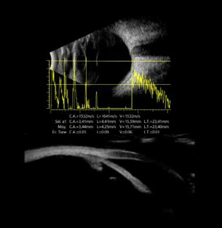
The ocular ultrasound is an examination that uses ultrasound waves to “see” inside the eye. It is particularly valuable when the fundus cannot be viewed, for example because of a dense cataract, intraocular hemorrhage, or corneal opacity. At Les Oliviers Eye Clinic in Sousse, Tunisia, ultrasound rapidly provides essential information about the retina, vitreous, and internal structures, helping guide diagnosis and treatment.
Ocular ultrasound
When is ocular ultrasound useful?
Ultrasound is a very effective alternative when slit-lamp examination or retinal photography cannot provide a clear view of the back of the eye. It is also used to confirm certain clinical suspicions and to prepare for surgery, particularly when vision is obscured by an opaque structure.
- Very dense cataract: Check the condition of the vitreous and retina before surgery.
- Vitreous hemorrhage: Examine the retina when it cannot be seen directly.
- Suspected retinal detachment: Help confirm or rule out the diagnosis.
- Eye trauma: Assess certain internal structures according to the circumstances.
How is the examination performed?
Ocular ultrasound is performed during a consultation or in a dedicated examination room. Depending on the protocol, a probe is placed gently on the closed eyelid or on the eye after anesthetic drops have been instilled, so the examination is painless. Gel may be used to improve ultrasound transmission. The doctor may ask you to look in different directions to examine several areas.
- Simple positioning: Sitting or lying down for a few minutes.
- Probe and gel: Gentle contact, with no radiation and no needle.
- Eye movements: To examine all internal areas.
- Immediate result: Interpretation is available on site according to the indication.
Is it painful?
No. Ocular ultrasound is generally painless. You may feel slight pressure on the eyelid, but the examination remains comfortable and brief.
What can be seen on ultrasound?
Ocular ultrasound makes it possible to examine the vitreous , the transparent gel inside the eye, the retina, and certain posterior structures. It can reveal membranes, traction, detachments, or internal opacities. In some cases, it can also help measure the length of the eye using an “A-scan” ultrasound, depending on the equipment and the assessment required.
- Vitreous: Opacities, hemorrhage, and membranes.
- Retina: Look for detachment or traction.
- Internal structures: Examine the back of the eye when the view is obscured.
- Measurements, when indicated: A-scan biometry in certain situations.
Practical advice before and after the examination
Ultrasound generally requires no preparation. Bring your medical documents, prescriptions, reports, and relevant history, and mention any severe pain or recent trauma. After the examination, you can resume your usual activities unless you are given specific instructions.
- Bring your previous examinations: OCT, fundus examination, and prescriptions if available.
- Tell us about pain or trauma: This helps determine urgency and the appropriate approach.
- Seek urgent care if you experience: sudden loss of vision, severe pain, flashes, and a dark curtain or shadow.
Frequently asked questions
Does ultrasound replace OCT?
No. OCT provides very detailed cross-sectional images of the retina, rather like microscopic slices, whereas ultrasound is especially useful when the view is obscured or when the vitreous and back of the eye need to be examined in a different way. The two examinations are complementary.
Do I need someone to accompany me?
Usually not. If other examinations involving dilation are planned on the same day, the team will advise you according to your comfort and whether you intend to drive.