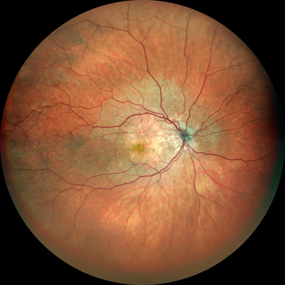
The retinal photography is a high-definition photograph of the fundus , including the retina, optic disc, and retinal blood vessels. It is a simple and rapid examination used to document the condition of the retina at a given moment and then compare images over time. At Les Oliviers Eye Clinic in Sousse, Tunisia, retinal photography is often used for screening and follow-up in conditions such as diabetes, AMD, hypertension, and glaucoma. It also helps explain findings to the patient using the image itself.
Retinal photography
What is retinal photography used for?
Retinal photography “freezes” a precise image of the fundus. It is useful for identifying lesions, confirming a clinical suspicion, and above all monitoring changes. It also improves communication, as viewing an image of the fundus often makes explanations clearer.
- Diabetic screening: Look for subtle abnormalities and provide regular follow-up.
- AMD and macular disease: Document the appearance of the fundus and compare it over time.
- Hypertension and blood vessels: Assess the appearance of retinal vessels.
- Optic nerve: Photographs of the optic disc are useful for glaucoma follow-up.
How is the examination performed?
You sit in front of a fundus camera with your chin on a support and your forehead against a bar. You are asked to look at a target while the device takes one or more photographs. Depending on your pupil and the purpose of the examination, dilation may be performed with eye drops to obtain clearer and wider images.
- Positioning: Chin and forehead stabilized while maintaining fixation.
- Brief flash: The light may be surprising, but it lasts only a moment.
- Several images: Sometimes central and peripheral images, depending on the indication.
- Dilation, sometimes: Blurred vision and light sensitivity for a few hours.
Is it painful?
No. Retinal photography is painless. The only possible discomfort is the camera flash, which may dazzle you for a few seconds.
What can be seen on a retinal photograph?
Retinal photography shows the appearance of the blood vessels, the macula, which is the central area of vision, the optic disc, which is the head of the optic nerve, and any visible fundus lesions. It also provides a visual record before and after treatment or simply throughout follow-up.
- Blood vessels: Caliber, abnormalities, and visible hemorrhages depending on the case.
- Macula: Deposits, changes, and visible signs depending on the condition.
- Optic disc: Appearance useful for monitoring the optic nerve.
- Comparison: The main advantage is being able to compare photographs over time.
Practical advice before and after
No special preparation is required. Bring your glasses and any previous examinations. If dilation is performed, bring sunglasses and avoid driving if your vision is affected.
- Bring your previous photographs: This makes comparison easier and more reliable.
- Sunglasses: Useful if dilation is planned.
- Tell us about your symptoms: Reduced vision, spots, or distortion help determine which images are needed.
Frequently asked questions
Is retinal photography sufficient for diabetes?
It is very useful for screening and follow-up. Depending on the findings, the doctor may recommend OCT to assess the macula or angiography to examine circulation and leakage.
Why are several photographs sometimes taken?
Because some lesions may be peripheral or because several areas need to be documented, such as the macula, optic disc, and vascular arcades, depending on your situation.