Back to top
+216 73 277 600
Examinations Surgery Technology Expertise

Optical Coherence Tomography (OCT)

The OCT (Optical Coherence Tomography) is an imaging examination that produces extremely fine cross-sectional images of the retina and/or optic nerve. It can be thought of as a scan, but one that uses light rather than radiation. The result is a detailed view of the retinal layers, helping detect abnormalities that may not be visible during a standard examination. At Les Oliviers Eye Clinic in Sousse, Tunisia, OCT is a central tool for diagnosing and monitoring macular diseases and glaucoma.

Purpose View the retina “in cross-section”
Principle Light-based imaging, without pain
Key point Highly precise and ideal for follow-up
What is Optical Coherence Tomography (OCT) used for?

OCT can detect very early retinal changes, assess tissue thickness, and monitor progress after treatment. It is particularly useful when a patient reports distorted vision or reduced visual acuity, or when the doctor is monitoring a chronic condition.

  • Macula: Edema, membranes, macular holes, and subtle abnormalities of the retinal layers.
  • AMD: Detection and monitoring of activity and follow-up after treatment.
  • Diabetes: Measurement of macular edema and monitoring of treatment response.
  • Glaucoma: Analysis of the optic nerve and certain nerve-fiber layers.
OCT is often complementary to other examinations such as fundus examination, angiography, and visual field testing. Each examination provides a different part of the picture.

How is the examination performed?

Optical Coherence Tomography is quick and painless. You place your chin on a support, look at a light, and the device scans the eye in a few seconds. There is no contact with the eye. Depending on the circumstances, the pupil may be dilated to improve image quality.

  • Positioning: Chin and forehead stabilized while looking at a target.
  • Rapid scan: A few seconds, without pain.
  • Cross-sectional images: The retina appears as a series of superimposed layers.
  • Dilation, sometimes: Depending on the eye and the image quality required.
Does OCT expose me to radiation?

No. OCT uses non-invasive light and does not use X-rays. It is a routine ophthalmic examination.

What does an OCT image show?

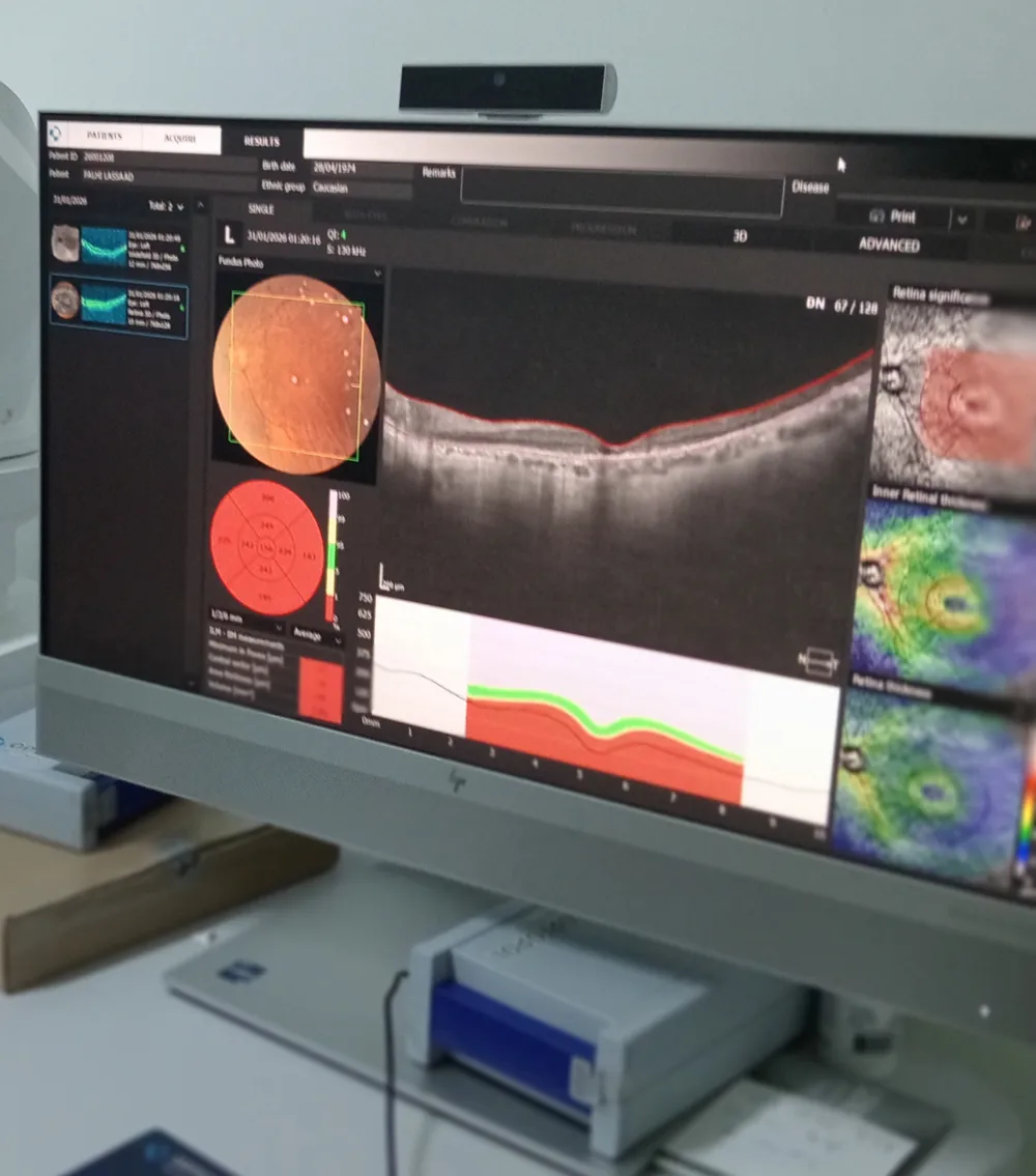
On OCT, the retina is shown in cross-section. Its layers can be distinguished, their thickness can be measured, and abnormalities such as fluid or edema, traction from a membrane, or changes in the macula can be identified. For glaucoma, OCT can assess the appearance of the optic nerve and the thickness of certain nerve-fiber layers.

  • Thickness: Measurements useful for diagnosis and follow-up.
  • Fluid / edema: Identification of pockets of fluid beneath or within the retina.
  • Traction: Membranes, vitreous adhesions, and subtle distortion.
  • Optic nerve: Helps with structural follow-up, particularly alongside visual field testing.
OCT optical coherence tomography in Sousse, Tunisia
A “normal” OCT is reassuring, but it must always be interpreted together with your symptoms and the clinical examination.

Practical advice before and after

OCT requires no special preparation. Bring your glasses and any previous examinations. If dilation is planned, vision may be blurred and light may be uncomfortable for a few hours. Bring sunglasses and avoid driving if you do not feel comfortable.

  • Bring your previous OCT scans: Comparison over time is very useful.
  • Blink naturally: A well-lubricated eye produces clearer images.
  • After dilation: Wear sunglasses and drive only if you feel comfortable.

Frequently asked questions

Why is OCT repeated regularly?

Because it allows changes to be monitored. Some diseases progress slowly, and OCT can detect renewed activity or stabilization, sometimes even before vision changes.

Does OCT replace angiography?

No. OCT shows structure, while angiography shows circulation and vascular leakage. Depending on the case, one examination, the other, or both may be used.