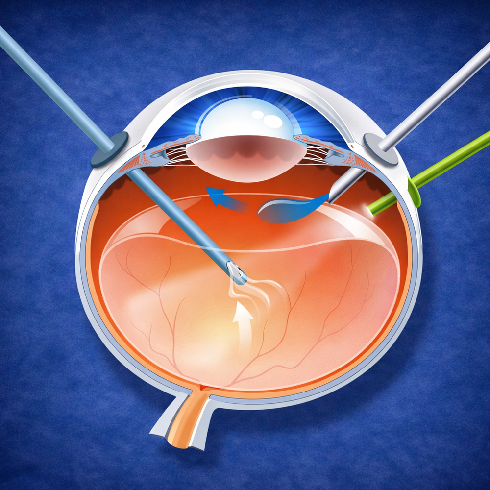
In retinal detachment, the retina separates from its supporting tissue like a film peeling away from a screen. vitrectomy is one of the main surgical techniques used to put it back in place. It makes it possible to remove traction that pulls on the retina, treat the tear or tears responsible, usually with laser, and then stabilize the retina with an internal tamponade such as gas or silicone oil while healing occurs. or only the affected layer . This tailored choice can make a major difference to visual recovery.
Vitrectomy | Retinal detachment
When is vitrectomy indicated?
In retinal detachment, the vitreous, the transparent internal gel, may pull on the retina, keep the tear open, or prevent proper reattachment. Vitrectomy allows treatment “from inside” the eye: the retina is freed, the tear is treated precisely, and the retina is placed in the best possible conditions for healing.
- Remove traction: The vitreous that is pulling on the retina is removed.
- Seal the tear: Laser is applied around the lesion to “weld” the retina in place.
- Reattach the retina: The retina is brought back into contact with its supporting tissue.
- Stabilize healing: An internal tamponade with gas or silicone is used depending on the situation.
How the operation is performed
Vitrectomy is carried out in the operating room under appropriate anesthesia. The surgeon uses micro-instruments through very small incisions. Once traction has been removed and the retina repositioned, laser treatment is applied around the tears. Finally, an internal tamponade may be used to keep the retina pressed in place during healing.
What the surgeon does
- Micro-incisions: Internal access using minimally invasive microsurgery.
- Vitrectomy: Removal of the vitreous and traction.
- Treatment of the tears: Laser treatment to reinforce the fragile area.
- Stabilization: Gas or silicone depending on the type of detachment.
Tamponade
In certain situations, a tamponade with gas or silicone oil is used to keep the retina or macula in the correct position while healing occurs.
- Gas: Gradually absorbs. Vision remains blurred while the bubble is present.
- Silicone oil: Provides longer stabilization. A later procedure to remove it may be necessary.
- Instructions: Air travel and high altitude are generally contraindicated while gas is present.
Why may positioning be required after surgery?
When gas is used, the bubble must press against the correct area, where the tear is located, to promote sealing. A particular posture, such as face down or with the head tilted, may be prescribed to optimize the effect of the tamponade.
Postoperative recovery
After vitrectomy, vision does not return “in one day.” It improves as healing progresses and depends greatly on whether the macula was involved. Eye drops are essential to reduce inflammation and protect the eye. Follow-up checks monitor pressure, the condition of the retina, and the position of the tamponade.
- Blurred vision at first: Very common, especially when gas is present.
- Close follow-up: Essential for a safe result.
- Protection and rest: According to instructions, avoid rubbing the eye and any physical effort that has not been authorized.
- Seek emergency care if you experience: a new curtain or shadow, significant pain, sudden loss of vision, or intense redness.
Frequently asked questions
Can the detachment recur?
Recurrence is possible, especially if new tears develop or scar tissue creates traction. Follow-up is used to detect any sign of fragility as early as possible.
Why is cataract sometimes discussed after vitrectomy?
In some patients, vitrectomy may accelerate clouding of the crystalline lens. If this occurs, cataract surgery may be discussed later.