Back to top
+216 73 277 600
Examinations Surgery Technology Expertise
Eye surgery Retinal detachment Retinal detachment Book an appointment

Retinal detachment

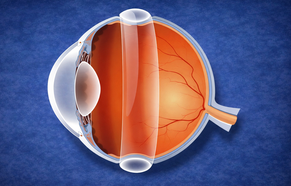
The retinal detachment occurs when the retina, the light-sensitive “film” at the back of the eye, separates from its supporting tissue. Without treatment, vision can deteriorate rapidly. The aim of surgery is to reattach the retina, treat the cause , which is often a retinal tear, and protect the macula, the central area responsible for fine vision. At Les Oliviers Eye Clinic in Sousse, Tunisia, two methods are used depending on your case: vitrectomy and scleral buckling.

Important to know: If you notice a “curtain” or rapid loss of vision, seek emergency care. The earlier the operation is performed, the better the chances of preserving useful vision.
Purpose Reattach the retina and treat the tear
Our techniques Vitrectomy and scleral buckling
Key point Emergency treatment + postoperative follow-up
Why is retinal detachment an emergency?

The retina depends on exchanges with the underlying tissues. When it detaches, it is no longer nourished properly. The longer the detachment persists, the more limited visual recovery may be. Timing is therefore important, especially if the macula is threatened or already involved.

Warning signs

  • Flashes of light: A sensation of flashes, especially in the dark.
  • A sudden shower of floaters: The abrupt appearance of numerous floating spots.
  • Shadow / curtain: A dark area advancing across the field of vision.
  • Loss of vision: Especially if central vision becomes distorted or decreases.

Our two techniques: vitrectomy and scleral buckling

The choice is not a matter of preference, but a medical decision based on the pattern of detachment, the presence of vitreous traction, the location of the tears, the condition of the crystalline lens, and the objective of long-term stability.

Vitrectomy

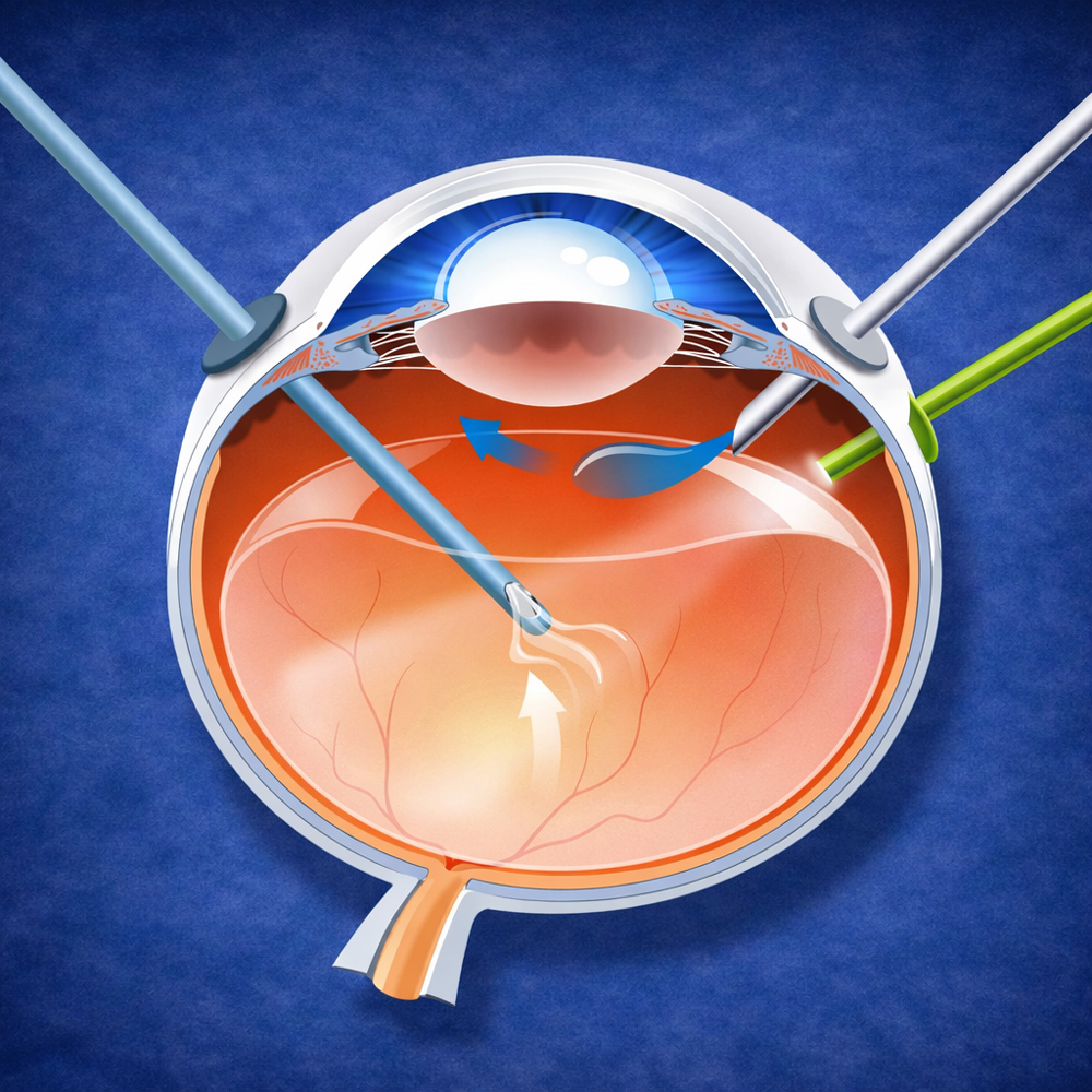
The vitrectomy consists of removing the vitreous gel, the transparent gel inside the eye, when it pulls on the retina or prevents proper reattachment. The surgeon then treats the tears, usually with laser, and may use an internal tamponade such as gas or silicone oil to hold the retina in place while it heals.

  • When? For complex detachments, traction, hemorrhage, and other situations depending on the indication.
  • Principle: Remove traction + seal the tear + stabilize the retina.
  • Afterwards: Specific positioning may be required depending on the tamponade used.
Vitrectomy in Sousse, Tunisia

Scleral buckling

Scleral buckling involves placing a silicone band or implant on the outer wall of the eye. This indents the wall inward, bringing it closer to the detached retinal area, reducing traction at the tear, and promoting reattachment.

  • When? For selected peripheral tears, depending on the patient’s profile and anatomy.
  • Principle: Bring the eye wall closer to the retina and relieve traction.
  • Follow-up: Regular checks confirm reattachment and monitor pressure.
Scleral buckling in Sousse, Tunisia
Can the two techniques be combined?

Yes. In certain situations, a combined approach may be considered to maximize stability, depending on the location of the tears, the degree of traction, and the complexity of the detachment.

Recovery and precautions: what really matters

After surgery, the eye needs time to heal. Vision improves gradually and depends particularly on whether the macula was involved, how long the retina was detached, and how healing progresses. Eye drops and follow-up appointments are essential because they help prevent inflammation, adjust pressure, and confirm that the retina remains attached.

  • Postoperative check-ups: They make the result safer and allow early detection of complications.
  • Eye drops: Anti-inflammatory drops, with or without antibiotics, according to your prescription.
  • Activities: Resume them gradually according to instructions, including any required positioning.
  • Seek emergency care again if you experience: a new shadow, significant pain, sudden loss of vision, or marked redness.

Frequently asked questions

Will vision return “as it was before”?

The first aim of surgery is to save the eye and preserve useful vision. Recovery depends greatly on the macula. If it was involved, vision may improve but remain less precise than before.

Why is positioning sometimes required?

Depending on the location of the tear and whether gas or silicone is used, maintaining a particular position may help keep the retina in place during the critical healing phase.

Should the other eye be monitored?

Depending on your risk factors, such as myopia, previous history, or peripheral retinal lesions, the other eye may require closer follow-up. Your ophthalmologist will advise you.